
In this lesson, we will use our own cell as an example to demonstrate the basic skills of installing slides (without bubbles).
Materials needed
a. Blank microscope slides and coverslips
b. Sterilized cotton head
c. tweezers
d. Methylene blue stain
Method of attaching a slide for a microscope
1. Clean the slides with Kim Whip paper.
2. Take a clean cotton swab (sealed package, sterile), rub the inside of the mouth gently.
3. Apply a cotton swab to the center of the slide for 2-3 seconds.
4. Add a drop of methylene blue solution to smear. Since methylene blue solution capable staining the cells with blue, easier to see under a microscope.
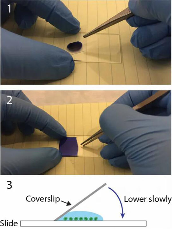
5. Place the coverslip on top.
Note: Lower the coverslip at an angle and slowly. Make sure that one side of the droplet touches the coverslip first. This allows the air to escape from the other side. Forceps can be used to help control coverslip.
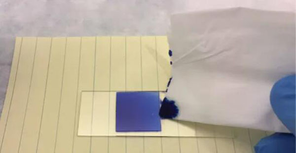
6. Use Kim Whip paper to touch one side of the coverslip to remove excess solution.
Note: For long-term storage, all edges of the coverslip can also be sealed with superglue (or nail polish).
Methylene blue
Methylene blue is a common alkaline staining that can be used to better view the nuclei were stained with animal cells. Methylene blue, and then bound to the very abundant DNA in the nucleus. Cells stained with methylene blue, shows the nucleus in dark blue. It also helps to display cells in the background. The shape of a cell helps to determine what the cell is (morphology).
Methylene blue can be replaced by most staining protocols that require Carmine or Janus Green B. Methylene blue is also used by lovers of the aquarium in order to prevent fungal diseases.
Wet mounting medium
Glycerol-based encapsulant, can provide a refractive index higher than that of pure water. You need to identify a specific cell structure.If you need a lower index of refraction, you can mix pure water with the glycerin solution to get the appropriate index of refraction.
When the glycerol concentration is high, it will also be a strong tendency for water from the sample is withdrawn. In particular, in the case of sensitive algae and other aquatic organisms to dehydration, there is a possibility that the specimen is deformed to contract. The glycerin mount can be sealed by applying superglue or nail polish to the sides of the cover glass. This keeps the cover glass in place for an extended period of time.
What will you see?
For methylene blue solution to stain the cell nucleus, the nucleus is in dark blue, the cytoplasm will find that it is colored in light blue.
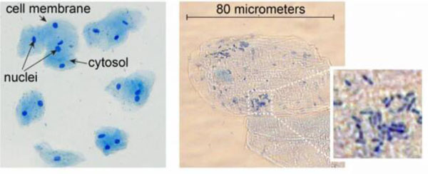
Also seen a small rod-shaped bacteria to the right of the image. Please do not worry, they are usually of oral microorganisms.
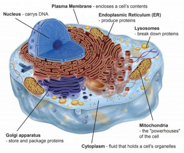
Animal cell anatomy
Cells are the “components” of life. The average male body contains about 30-40 trillion cells. The diameter of the cell is only about 20 micrometers (one millionth of a meter), but it’s still surprisingly complex.Each organelle has its own features.
Post time: Oct-26-2021


