
Nature: Smart microscope slides can detect cancer
October 7, 2021/Bio Valley BIOON/—In a new study, Associate Professor Belinda S. Parker and Professor Brian Abbey of La Trobe University, Australia, and their team discovered that by modifying traditional microscopes on the nanoscale The surface of the glass slide, biological structure and cells will show amazing color contrast, which can be used to detect diseases instantly. The relevant research results will be published in the journal Nature on October 7, 2021. The title of the paper is “Colorimet
October 7, 2021/Bio Valley BIOON/—In a new study, Associate Professor Belinda S. Parker and Professor Brian Abbey of La Trobe University, Australia, and their team discovered that by modifying traditional microscopes on the nanoscale The surface of the glass slide, biological structure and cells will show amazing color contrast, which can be used to detect diseases instantly. The relevant research results were published in Nature on October 7, 2021. The title of the paper is “Colorimetric histology using plasmonically active microscope slides”.
Professor Abbey has developed this technology at La Trobe University in the past five years with co-inventor Dr. Eugeniu Balaur. Professor Abbey said, “Current tissue imaging methods often rely on staining or marking cells to make them visible under the microscope. Even with staining or marking, it is still challenging for pathologists to detect cancer cells. Some samples may be misdiagnosed, especially in the very early stages of the disease. Recent breakthroughs in nanotechnology have allowed us to manipulate the interaction of light with biological tissues so that abnormal cells appear to have a different color from healthy cells. The image on the slide is compared with the traditional staining method, just like watching a color TV, but before, I saw black and white TV pictures.”
In this new research, Professor Abbey’s team and Associate Professor Parker’s team have tried to use this new technology called NanoMslide as an auxiliary method for diagnosing very early breast cancer.
Associate Professor Parker said that current technology can make it difficult to distinguish early forms of breast cancer from benign lesions, especially when there are not many abnormally shaped cells in complex tissues. NanoMslide makes this diagnosis easier.
Associate Professor Parker said, “When I observed the tissues under the microscope of NanoMslide for the first time, I was incredibly excited. For the first time I saw cancer cells suddenly appear in front of me. Their color is different from the surrounding tissues and they are very easy. Distinguish them from the surrounding cells.”
Associate Professor Parker believes that NanoMslide will complement the existing stains currently in use to achieve a more consistent cancer diagnosis. “Based on our preliminary research results on NanoMslide, we believe that this platform is really useful in the diagnosis of early breast cancer, and in other cancers, we really just want to single out some cancer cells in complex tissues or blood samples. ”
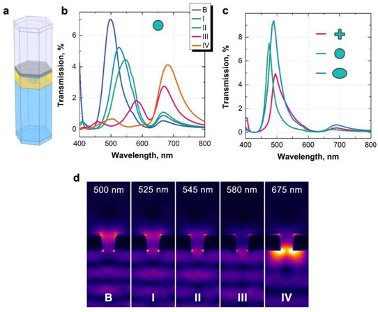
Finite element method (FEM) optical transmission simulation, picture from Nature, 2021, doi:10.1038/s41586-021-03835-2.
Professor Abbey’s team developed their glass slide technology by leveraging the open access equipment and expertise provided by the Melbourne Nanomanufacturing Centre. The team will work with the Melbourne Nanofabrication Center and the Australian National Fabrication Facility (ANFF) network to begin mass production of their slides to enter the market and solve a wide range of medical and non-medical imaging problems.
Professor John Dewar AO, Vice-Chancellor of La Trobe University, said that the invention of NanoMslide and its application in improving cancer diagnosis highlights the important role that universities like La Trobe University play in research innovation, and research innovation can improve The power of life.
Professor Dewar said, “As this outstanding invention transforms from an outstanding concept into a potentially life-saving solution, La Trobe University has proven that it can be achieved when outstanding research innovation is combined with a strong industry partner. What kind of results.”
Reference materials:
Brian Abbey et al. Colorimetric histology using plasmonically active microscope slides. Nature, 2021, doi:10.1038/s41586-021-03835-2.
Post time: Dec-02-2021


