
What Is Microbiology?
Microbiology is the scientific study of microorganisms, including bacteria, fungi, protozoa, algae, and other microscopic organisms. Because many microorganisms cannot be seen clearly with the naked eye, laboratory microscopy plays an important role in observing their morphology, structure, distribution, and interaction with biological samples.
In clinical laboratories, research institutions, food testing laboratories, pharmaceutical companies, environmental laboratories, and veterinary facilities, microbiology workflows often involve collecting, preparing, staining, and examining samples under a microscope.
A microscope slide is therefore a basic but important consumable in microbiological testing. The quality of the glass surface, optical clarity, cleanliness, dimensional accuracy, and sample adhesion can all influence the efficiency and reliability of microscopic observation.
For laboratories and distributors purchasing laboratory consumables in volume, selecting the appropriate slides from a professional microscope slides manufacturer can help maintain consistent sample preparation and testing performance.
The Workflow Of the Typical Microbiology
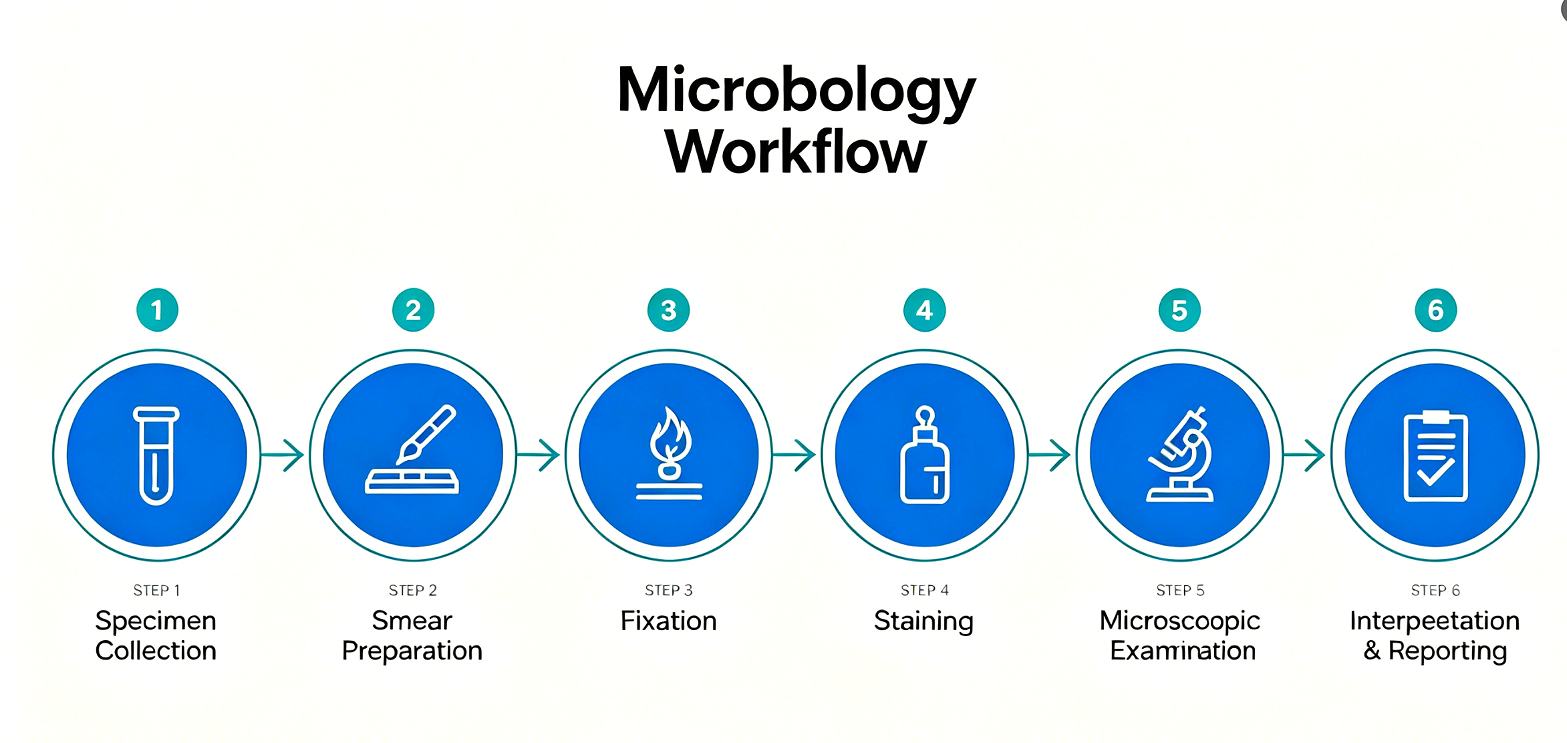
Although microbiology procedures vary according to the application, a typical workflow can include several key stages.
Step 1: Sample Collection
The laboratory first collects a biological or environmental sample. Depending on the application, the sample may come from clinical specimens, food, water, soil, cultures, or other sources.
Proper sample collection and handling are essential because contamination or improper storage can affect subsequent analysis.
Step 2: Sample Preparation
The collected sample is prepared for microscopic examination. A small amount of material may be transferred onto a microscope slide and spread into a thin layer.
The purpose is to create a suitable sample thickness for light to pass through the specimen and allow microscopic structures to be observed.
Step 3: Fixation
In many microbiological applications, the sample is fixed to the glass surface. Fixation helps preserve cellular or microbial structures and improves the stability of the specimen during subsequent staining and washing procedures.
This is one reason why surface properties of microscope slides are important.
Step 4: Staining
Some microorganisms and cellular structures are difficult to distinguish under a microscope without staining.
Common microbiological staining procedures include Gram staining and other differential or special staining techniques. Staining can increase contrast between microorganisms and the surrounding background, making important morphological characteristics easier to observe.
Step 5: Microscopic Examination
The prepared slide is examined under a microscope. Laboratory personnel may evaluate characteristics such as cell shape, size, arrangement, staining response, and the presence of particular structures.
Step 6: Interpretation and Reporting
The microscopic observations are combined with other laboratory information to support identification, research, quality control, or diagnostic decisions.
Therefore, the microscope slide is not simply a piece of glass. It is part of the sample preparation system connecting the specimen to the microscope.
Why Are Microscope Slides Used in Microbiology?
The primary function of a microscope slide is to provide a stable, planar, optically uniform platform that positions the specimen within the focal plane of the microscope. Four functional roles distinguish a high-quality slide from an inferior one:
● Specimen Support and Positioning
Microorganisms and biological specimens must be immobilized in a controlled, repeatable plane to enable consistent focusing and examination. Standardized microscope slide dimensions (typically 25 × 75 mm or 25.4 × 76.2 mm, 1.0–1.2 mm thickness) ensure compatibility with universal microscope stages, mechanical stage clips, and automated slide-handling systems.
● Specimen Retention During Processing
During fixation, staining, decolorization, and rinsing, specimens — particularly delicate cytological preparations, thin tissue sections, and dilute microbial smears — are prone to detachment from conventional untreated glass surfaces. Adhesive microscope slides address this limitation through surface-modified chemistries that promote stronger specimen–glass binding, reducing sample loss and the need for repeated preparation.
● Optical Clarity and Imaging Consistency
High-quality super white glass microscope slides offer superior light transmission, low autofluorescence, and minimal optical distortion compared with standard soda-lime glass. Consistent thickness, flatness, and surface smoothness ensure that imaging conditions remain reproducible across slides, batches, and operators — a requirement for quantitative microscopy and automated image analysis.
● Workflow Efficiency and Throughput
When specimens detach during processing, technicians must repeat smear preparation, fixation, and staining — consuming reagents, technician time, and potentially irreplaceable clinical material. Adhesive microscope slides and positive charged glass slides reduce rework rates, improve workflow consistency, and increase effective laboratory throughput. For high-volume laboratories and distributors, sourcing adhesive microscope slides wholesale directly from a manufacturer is a cost-effective strategy when sample retention is a critical quality parameter.
Why Choose Positive Charged Glass Slides?
For applications where specimen adhesion is a critical success factor, positive charged glass slides are a well-established solution. A positive charged glass slides manufacturer produces slides whose surfaces are chemically modified — typically through silanization or amino-functionalization — to introduce a net positive electrostatic charge.
Because most biological materials — including bacterial cell walls, nucleic acids, and many cellular proteins — carry a net negative charge at physiological pH, the electrostatic attraction between the negatively charged specimen and the positively charged slide surface significantly enhances binding affinity.
Jiangsu Huida Microscope Slides for B2B Microbiology Applications
For laboratories, distributors, and international buyers seeking a reliable source of microbiology consumables, Jiangsu Huida Medical Instruments Co., Ltd. manufactures a comprehensive range of microscope slides and associated products designed for professional B2B applications.
Within microbiology and related laboratory workflows, adhesive microscope slides and positive charged glass slides are particularly relevant when specimen retention is a priority. Jiangsu Huida's product portfolio includes:
● Plain Microscope Slides (7101/7102 series) — manufactured from soda-lime or super white glass, pre-cleaned and ready for routine microbiology, hematology, and educational applications
● Color Frosted Microscope Slides — featuring color-coded frosted marking areas for rapid specimen identification and laboratory workflow organization
● Adhesive Microscope Slides — surface-treated for enhanced specimen retention during multi-step staining and processing
● Positive Charged Glass Slides — amino-silanized for strong electrostatic binding of negatively charged biological specimens, suitable for IHC, ISH, FISH, and cytology
● Cover Glass / Cover Slips — available in standard thickness grades (#1.0, #1.5, #2.0) and multiple dimensions, compatible with manual and automated coverslipping systems.
FAQ
1. What are adhesive microscope slides used for?
Adhesive microscope slides are surface-treated to improve the attachment of biological specimens to the glass surface. They are essential when specimens must remain adherent through fixation, staining, washing, decolorization, or other multi-step laboratory processing — particularly in IHC, ISH, FISH, and cytology applications.
2. What is the difference between positive charged glass slides and plain microscope slides?
Positive charged glass slides are chemically modified (typically via amino-silanization) to introduce a net positive surface charge, which electrostatically attracts negatively charged biological materials. Plain microscope slides have an untreated or minimally treated glass surface and are suitable for routine applications where enhanced adhesion is not required.
3. Can microscope slides be used for fluorescence microscopy?
Yes, but glass selection is important. Standard soda-lime glass may exhibit autofluorescence at certain excitation wavelengths. For fluorescence applications, super white glass microscope slides with low autofluorescence specifications are recommended.
4. Where can I buy adhesive microscope slides wholesale?
Professional laboratories, distributors, importers, and medical supply companies can source adhesive microscope slides wholesale directly from manufacturers. When evaluating a supplier, consider glass quality, surface treatment consistency, dimensional tolerance, packaging options, customization capability, production capacity, and quality certifications.
5. Are cover glass products available alongside slides?
Yes. A complete microscopy consumables supplier typically offers both microscope slides and matching cover glass / cover slips in standard thickness grades and dimensions, ensuring compatibility across the specimen preparation workflow.
Post time:2026-08-27


